How Retinal Imaging Revolutionizes the Management of Glaucoma

How Retinal Imaging Revolutionizes the Management of Glaucoma
Glaucoma is a leading cause of irreversible blindness, affecting millions worldwide. This silent thief of sight often progresses without symptoms until significant vision loss occurs. However, advancements in retinal imaging have revolutionized how glaucoma is detected, monitored, and managed, allowing for earlier intervention and better outcomes for patients.
Understanding Glaucoma and Its Challenges
Glaucoma is characterized by damage to the optic nerve, often due to elevated intraocular pressure (IOP). This damage can lead to peripheral vision loss and, eventually, total blindness if left untreated. One of the greatest challenges in managing glaucoma is that the disease can progress silently, with no noticeable symptoms in its early stages. This makes regular eye exams and advanced diagnostic tools, such as retinal imaging, critical in preserving vision.
What Is Retinal Imaging?
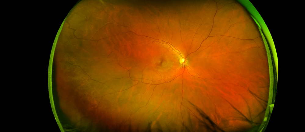
Retinal imaging involves the use of advanced technology to capture high-resolution images of the retina, optic nerve, and surrounding structures. These images provide an in-depth look at the health of the eye and allow optometrists to detect changes that might indicate glaucoma or other eye conditions.
Common types of retinal imaging used in glaucoma management include:
Optical Coherence Tomography (OCT): A non-invasive imaging test that uses light waves to create cross-sectional images of the retina. OCT is invaluable in measuring the thickness of the retinal nerve fiber layer (RNFL), a key indicator of glaucoma progression.
Fundus Photography: A digital imaging technique that captures detailed photographs of the retina and optic nerve head. These images help in monitoring structural changes over time.
Scanning Laser Ophthalmoscopy (SLO): A method that provides three-dimensional views of the optic nerve and retina for comprehensive analysis.
Early Detection and Diagnosis
Retinal imaging plays a crucial role in the early detection of glaucoma. By capturing detailed images of the optic nerve and retinal layers, optometrists can identify subtle changes that might indicate early-stage glaucoma. This allows for earlier intervention, potentially slowing disease progression and preserving vision.
Monitoring Disease Progression
For patients already diagnosed with glaucoma, retinal imaging provides a reliable way to monitor disease progression. Regular imaging allows optometrists to compare current and past images, tracking changes in the optic nerve and retinal nerve fiber layer. This data helps in determining the effectiveness of treatment plans and making necessary adjustments.
Retinal imaging is not only beneficial for diagnostic and monitoring purposes but also for educating patients. Visualizing the damage caused by glaucoma or observing the stability of the optic nerve over time can help patients better understand their condition and the importance of adhering to treatment plans.
Personalized Treatment Plans
Advanced retinal imaging technology enables optometrists to tailor treatment plans to each patient's unique needs. By closely monitoring changes in the optic nerve and retinal structures, eye care professionals can adjust medications, recommend surgical options, or explore other interventions to manage the disease effectively.
Schedule Your Comprehensive Eye Exam Today
At Texas State Optical, we are committed to providing state-of-the-art eye care to our patients. Our use of advanced retinal imaging technology ensures that glaucoma and other eye conditions are detected and managed with precision. Whether you’re due for a routine eye exam or need specialized glaucoma care, we are here to help.
Glaucoma may be silent, but with the help of advanced retinal imaging, it doesn’t have to rob you of your vision. Contact Texas State Optical to schedule your comprehensive eye exam and take the first step toward protecting your sight. Visit our office in Buda, Texas, or call (512) 991-8656 to book an appointment today.


